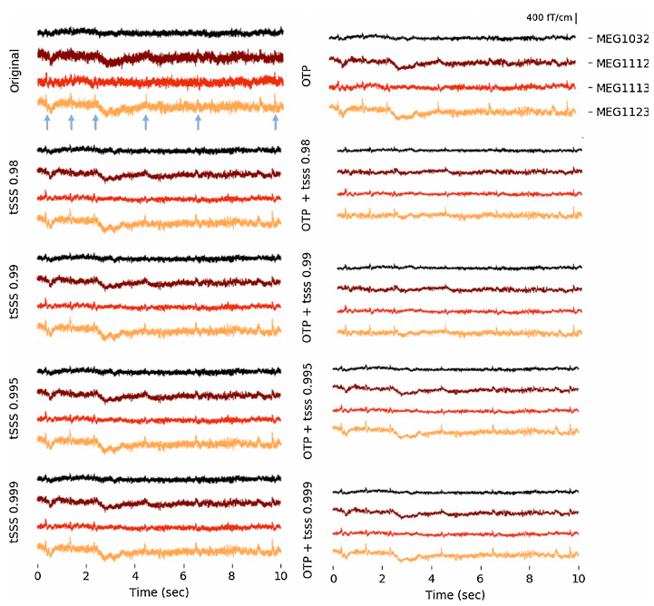
This is done by applying a time-domain, cross-validation procedure in which the signal of each channel, one by one, is compared to the signals of all other channels.
The purpose of this comparison is to find the signal component of each channel that is not consistent with the information of the other channels. Thus, the extracted signal component can be assumed to mainly consist of individual sensor noise and artifacts, and, therefore, we can remove this signal component from the original signal.
Now we are left with data that has less sensor noise than the original data.
The effect of Sig3 Denoise1™ is most significant at relatively high frequencies (above the power line frequency) where there is usually not much external interference and the sensor noise dominates the spectrum.
Many clinicians believe that the precursor of epileptic seizures occurs at high (gamma) frequencies, and these weak high-frequency oscillations (HFO) are difficult to detect due to sensor noise. With Sig3 Denoise1™ , this task should be much easier, which could be very important in terms of finding the true location of epileptic foci in the brain.
Please Note: This product shown does NOT have FDA clearance.
References
- Clarke, M., Larson, E., Tavabi, K., and Taulu, S. (2020). Effectively combining temporal projection noise suppression methods in magnetoencephalography. Journal of Neuroscience Methods 108700.
- Larson, E., and Taulu, S. (2018). Reducing Sensor Noise in MEG and EEG Recordings Using Oversampled Temporal Projection. IEEE Transactions on Biomedical Engineering 65, 1002–1013.
- Yumoto, E. Larson, S. Taulu, “Validation of MEG sensor noise reduction by Oversampled Temporal Projection” Japan Biomag Poster P07 (2019)
